Scanner and dental imaging
TIA dentico, Belgrade - Scanner and dental imaging
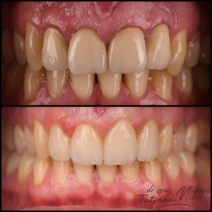
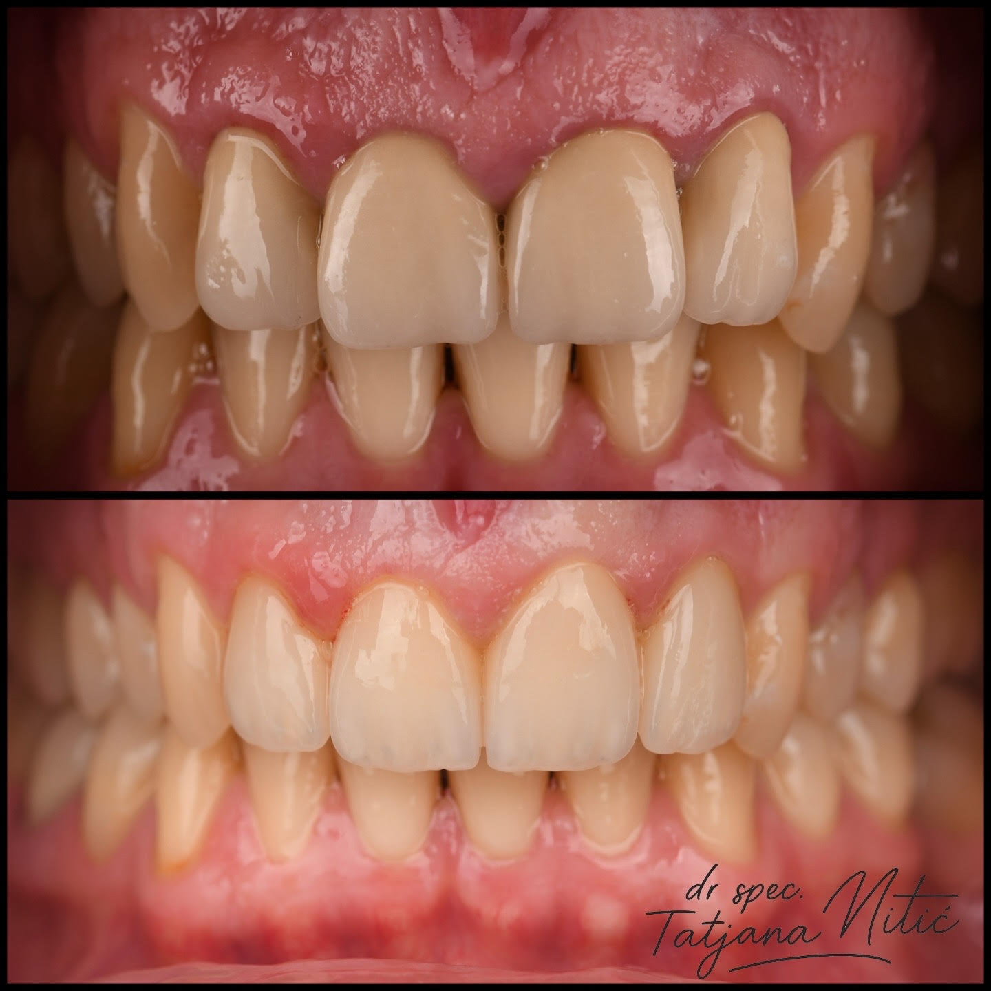
Dental Imaging – A Key Part of Diagnosis and Endodontic Treatment
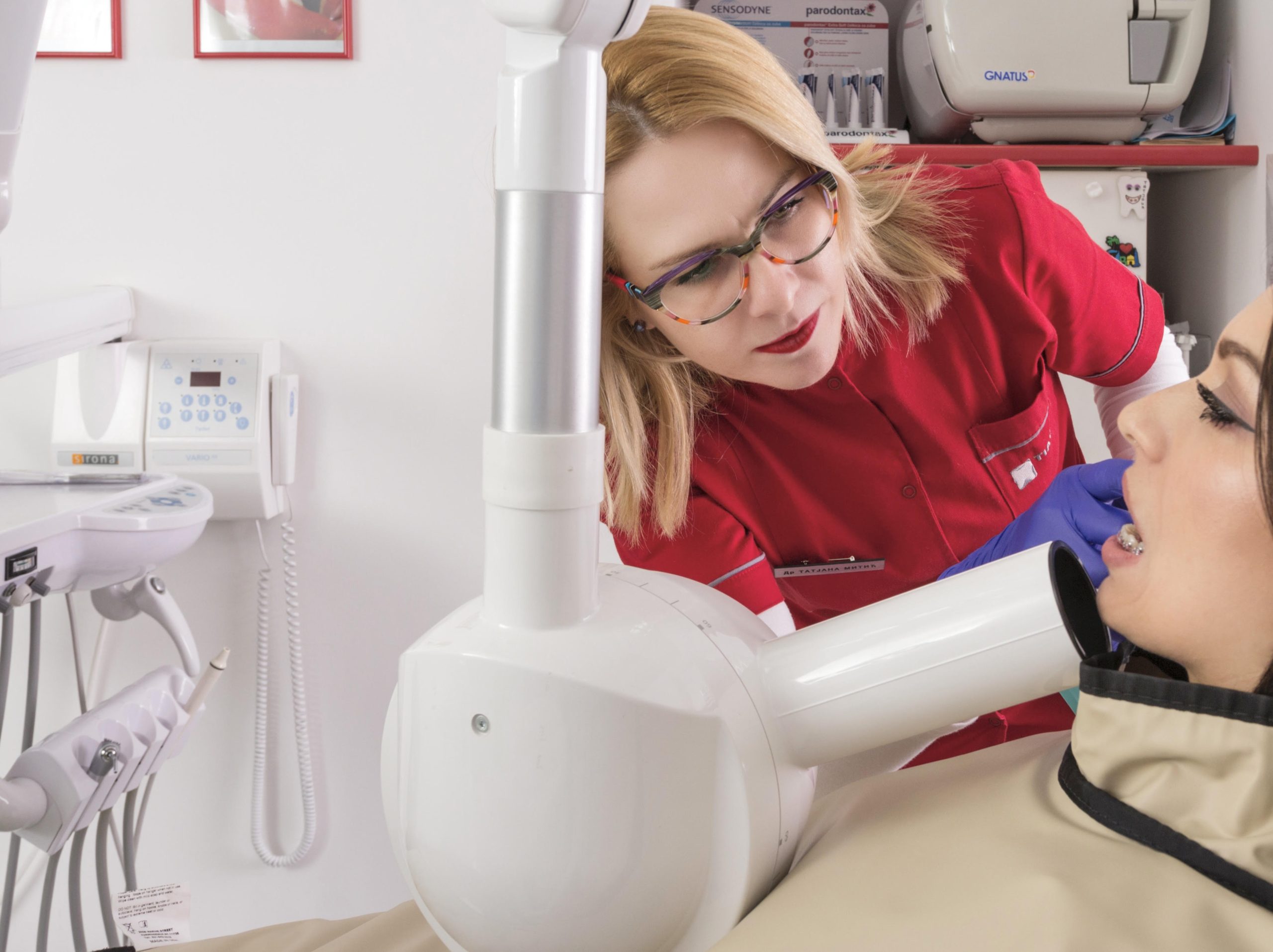
At our clinic, precise diagnostics form the foundation of every successful treatment procedure. That’s why dental imaging plays a crucial role in our workflow, especially when it comes to endodontic (root canal) treatment.
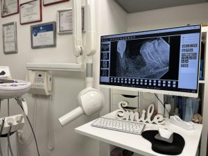
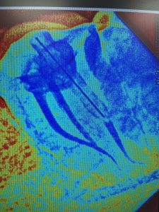
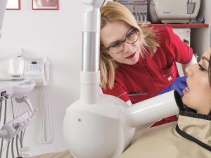
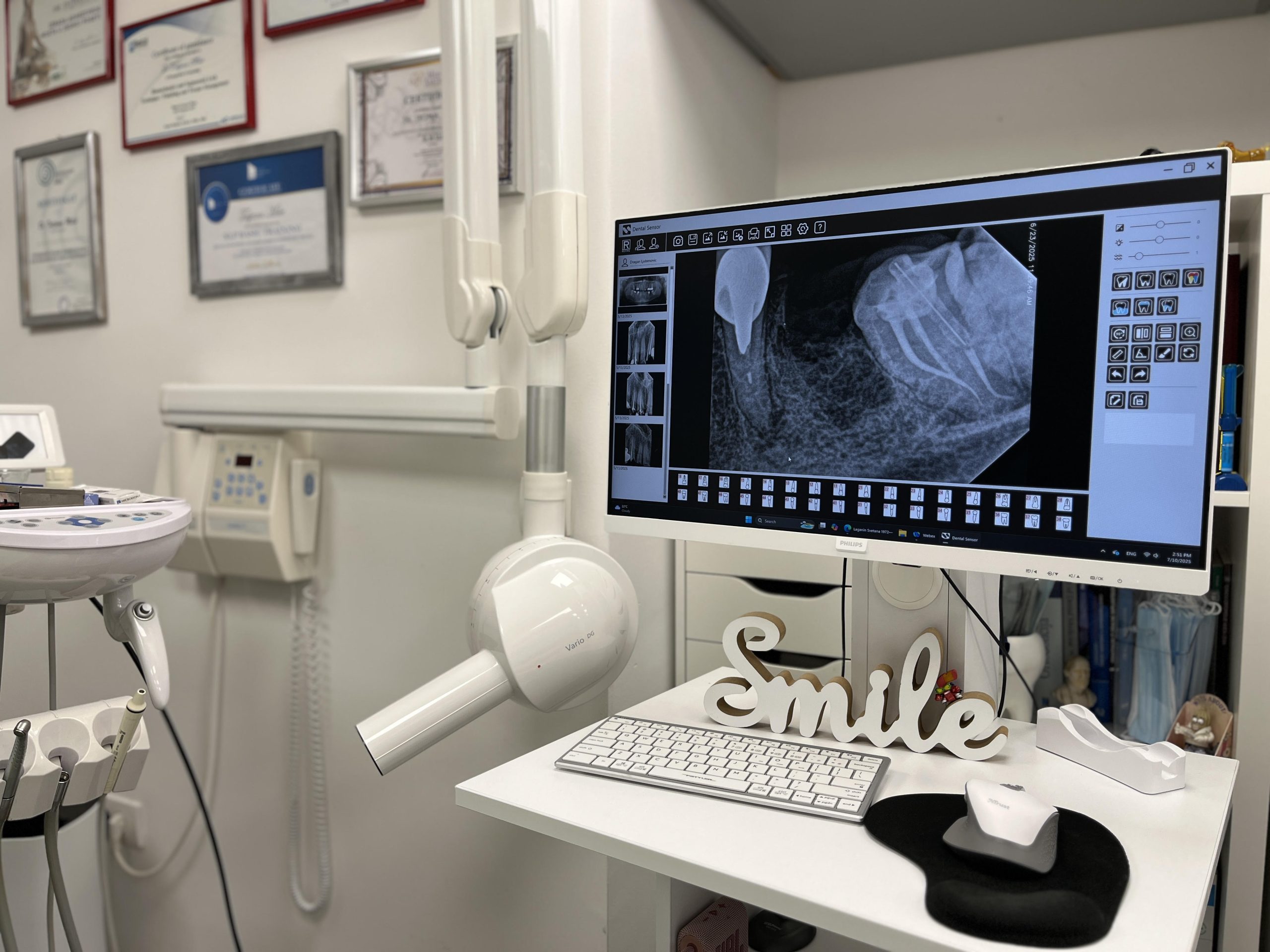
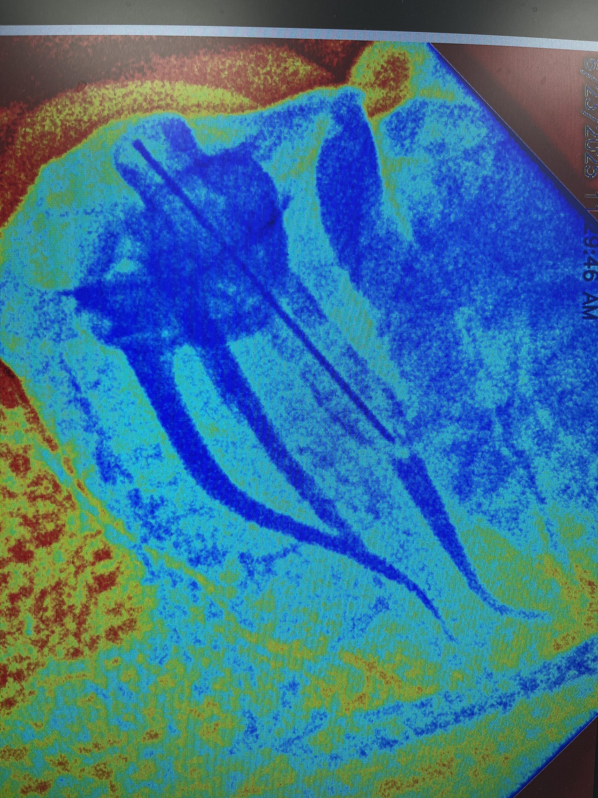
We use a modern digital intraoral device for retroalveolar imaging, which provides a detailed view of the tooth root, periapical area, and structures not visible during a clinical examination. This type of imaging offers a high level of accuracy with minimal radiation exposure, ensuring maximum safety and comfort for the patient.
Retroalveolar X-rays Are Essential For:
- Establishing an accurate diagnosis of the tooth and surrounding tissues
- Planning endodontic therapy
- Monitoring treatment progress
- Controlling the quality of canal filling
- Long-term evaluation of treatment success
Digital technology allows for instant image display, magnification, and real-time analysis, significantly improving diagnostic accuracy and treatment outcomes.
By integrating this system into daily practice, we achieve a high level of control and safety—for both the practitioner and the patient.
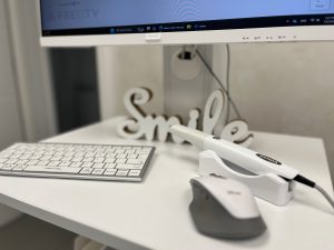
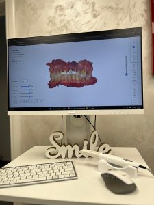
In recent years, technological advancements have increasingly led us toward a fully digital protocol. Intraoral scanners and digital impressions are becoming a regular part of everyday dental practice.
This raises the question: What are the advantages of digital impressions compared to conventional impression techniques?
According to S. Sharma and colleagues, the main issues with analog impressions include:
- Increased gag reflex
- Poor adhesion between impression material and tray
- Longer working time
- Limited mouth opening
- Material shrinkage
- Plaster contraction during model casting
On the other hand, researchers from Harvard have shown that 89% of impressions in fixed prosthodontics contain measurable errors.
In contrast, manufacturers of intraoral scanners highlight several benefits of digital impressions, including:
- Faster scanning
- Greater patient comfort
- Improved communication
- Cleaner workflow
- Higher precision
- Better fit of prosthetic restorations
From a clinical perspective, comparing the average time for conventional impressions (around 10 minutes) with intraoral scanning (around 4 minutes), it becomes clear that intraoral scanners offer a significant advantage.
When it comes to patient comfort, digital technology again takes the lead. A study by Yuzbasioglu and colleagues showed that 100% of participants preferred digital impressions over conventional methods.